Home » Without Label » Rib Cage Anatomy : The Thoracic Cage Anatomy And Physiology I : Together with the sternum, thoracic vertebrae, and costal cartilages, the ribs form the thoracic cage, also called the bony thorax.
Rib Cage Anatomy : The Thoracic Cage Anatomy And Physiology I : Together with the sternum, thoracic vertebrae, and costal cartilages, the ribs form the thoracic cage, also called the bony thorax.
Rib Cage Anatomy : The Thoracic Cage Anatomy And Physiology I : Together with the sternum, thoracic vertebrae, and costal cartilages, the ribs form the thoracic cage, also called the bony thorax.. The final two pairs of ribs are floating ribs and the cartilage of these ribs tends to end within the abdominal musculature. Internal intercostals can be found in the layer of muscles directly below the external intercostals. However, only seven have a direct articulation with the sternum. There are twelve pairs of ribs, all of which articulate with the vertebral column. Related posts of rib cage diagram with organs womens body parts stomach.
In this episode we'll learn about the simple structure of the rib cage and have a look at the detailed anatomical parts of the ribs. This packet goes over the gross anatomy of the sternum and rib cage. The ribs are a set of twelve paired bones which form the protective 'cage' of the thorax. There are two sets of intercostal muscles within the rib cage. All these 12 pairs are attached to the spines with their two attachment sites.
3d Illustration Of Human Skeleton System Rib Cage With Labels Anatomy Anterior View Canstock from comps.canstockphoto.com Rib cage, in vertebrate anatomy, basketlike skeletal structure that forms the chest, or thorax, and is made up of the ribs and their corresponding attachments to the sternum (breastbone) and the vertebral column. External intercostals can be found on the surface, just below the skin, and provide muscle contractions which assist with inhalation. Rib cage anatomy in homo erectus suggests a recent evolutionary origin of modern human body shape. At the chest, many rib bones connect to the sternum via costal cartilage,. Introduction to the structure of the ribcage and ribs: In this episode we'll learn about the simple structure of the rib cage and have a look at the detailed anatomical parts of the ribs. Lateral view of a pair of ribs articulating with the thoracic vertebrae. This packet goes over the gross anatomy of the sternum and rib cage.
Rib cage pain can be caused.
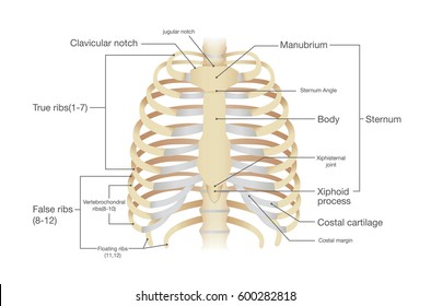
The thoracic cage consists of the 12 thoracic vertebrae, the associated intervertebral discs, 12 pairs of ribs with their costal cartilages, and the sternum. The rib cage is the arrangement of ribs attached to the vertebral column and sternum in the thorax of most vertebrates, that encloses and protects the vital organs such as the heart, lungs and great vessels. We examined the thoracic vertebrae last lab, so here we will only examine the ribs and sternum. There are 12 pairs of ribs. Related posts of rib cage diagram with organs womens body parts stomach. They are extremely light, but highly resilient; The upper edge is round and the lower sharp. The ribs are a set of twelve paired bones which form the protective 'cage' of the thorax. It discusses the specific anatomy of the ribs and costal cartilages, along with the sternum. Lateral view of a pair of ribs articulating with the thoracic vertebrae. Womens body parts stomach 4 photos of the womens body parts stomach body diagram stomach, body parts digestive system, body parts in stomach area, body parts liver, body parts spleen, human body parts stomach, woman body organs, woman body parts found, stomach, body diagram stomach, body parts digestive system, body. Lewis (1918) gray's anatomy 20th ed (in public domain at yahoo or bartleby) images: It's designed to move with respiration, the ribs rising and lowering with each breath, thus increasing the capacity of the chest cavity while reducing its pressure.
The costocorporeal joint is where the rib head connects with two adjacent vertebral bodies and the disc between them. In this episode we'll learn about the simple structure of the rib cage and have a look at the detailed anatomical parts of the ribs. The bottom of the vertebral body above has a costal demifacet (partial facet), and so does the top of the vertebral body below. They are extremely light, but highly resilient; The top edge of the manubrium has a depression called the suprasternal or jugular notch.
The Anatomy Of The Laboratory Mouse from www.informatics.jax.org The bones of the rib cage are the sternum, the 12 thoracic vertebrae and the 12 pairs of ribs. Related links to external sites (from bing) these images are a random sampling from a bing search on the term rib cage anatomy. Anatomy of right side of back of rib cage / rib cage wikipedia.the rib cage is the part of the axial skeleton that protects the vital organs within the thoracic (chest) it is made up of the ribs that articulate at the back with the vertebral column (vertebrae) and at the there are 12 ribs on each side (left and right) and a clavicle (collarbone) on the left and right as well. Introduction to the structure of the ribcage and ribs: The rib cage is the arrangement of ribs attached to the vertebral column and sternum in the thorax of most vertebrates, that encloses and protects the vital organs such as the heart, lungs and great vessels. They are extremely light, but highly resilient; Lateral view of a pair of ribs articulating with the thoracic vertebrae. The ribs are curved, flat bones which form the majority of the thoracic cage.
Anatomy the rib cage is a bony structure found in the chest (thoracic cavity).
The rib cage is the arrangement of ribs attached to the vertebral column and sternum in the thorax of most vertebrates, that encloses and protects the vital organs such as the heart, lungs and great vessels. The thoracic cage consists of the 12 thoracic vertebrae, the associated intervertebral discs, 12 pairs of ribs with their costal cartilages, and the sternum. Lewis (1918) gray's anatomy 20th ed (in public domain at yahoo or bartleby) images: They are extremely light, but highly resilient; They articulate with the vertebral column posteriorly, and terminate anteriorly as cartilage (known as costal cartilage). The costocorporeal joint is where the rib head connects with two adjacent vertebral bodies and the disc between them. It is made up of 12 pairs of ribs. The bones of the rib cage are the sternum, the 12 thoracic vertebrae and the 12 pairs of ribs. It may occur after an obvious injury or without explanation. There are twelve (12) pairs of ribs and all articulate posteriorly with the thoracic vertebrae. The thoracic cage surrounds and protects the heart and lungs in the thoracic cavity. Instead, anatomists classify the ribs as flat bones, and they are located within the axial skeleton. This packet goes over the gross anatomy of the sternum and rib cage.
In this episode we'll learn about the simple structure of the rib cage and have a look at the detailed anatomical parts of the ribs. Anatomy the rib cage is a bony structure found in the chest (thoracic cavity). Related posts of rib cage diagram with organs womens body parts stomach. The ribs are curved, flat bones which form the majority of the thoracic cage. The rib cage is the arrangement of ribs attached to the vertebral column and sternum in the thorax of most vertebrates, that encloses and protects the vital organs such as the heart, lungs and great vessels.
Rib Cage Anatomy Images Stock Photos Vectors Shutterstock from image.shutterstock.com There are twelve pairs of ribs. Rib cage anatomy the rib cage, shaped in a mild cone shape and more flexible than most bone sets, is made up of varying elements such as the thoracic vertebra, 12 equally paired ribs, costal cartilage, and held together anteriorly by the sternum. The costocorporeal joint is where the rib head connects with two adjacent vertebral bodies and the disc between them. The bones of the rib cage are the sternum, the 12 thoracic vertebrae and the 12 pairs of ribs. Basic anatomy of the rib cage. It's designed to move with respiration, the ribs rising and lowering with each breath, thus increasing the capacity of the chest cavity while reducing its pressure. Lateral view of a pair of ribs articulating with the thoracic vertebrae. On the interior wall of the rib body is a channel, sulcus costae, with blood vessels and nerves.
Internal intercostals can be found in the layer of muscles directly below the external intercostals.
External intercostals can be found on the surface, just below the skin, and provide muscle contractions which assist with inhalation. The thoracic cage surrounds and protects the heart and lungs in the thoracic cavity. The costocorporeal joint is where the rib head connects with two adjacent vertebral bodies and the disc between them. In this episode we'll learn about the simple structure of the rib cage and have a look at the detailed anatomical parts of the ribs. They articulate with the vertebral column posteriorly, and terminate anteriorly as cartilage (known as costal cartilage). Basic anatomy of the rib cage. A rib has a flat body, as you can see from the picture of the anatomy of the human rib cage. Internal intercostals can be found in the layer of muscles directly below the external intercostals. It is made up of 12 pairs of ribs. Lewis (1918) gray's anatomy 20th ed (in public domain at yahoo or bartleby) images: Lateral view of a pair of ribs articulating with the thoracic vertebrae. The ribs are curved, flat bones which form the majority of the thoracic cage. Rib cage pain may be sharp, dull, or achy and felt at or below the chest or above the navel on either side.